
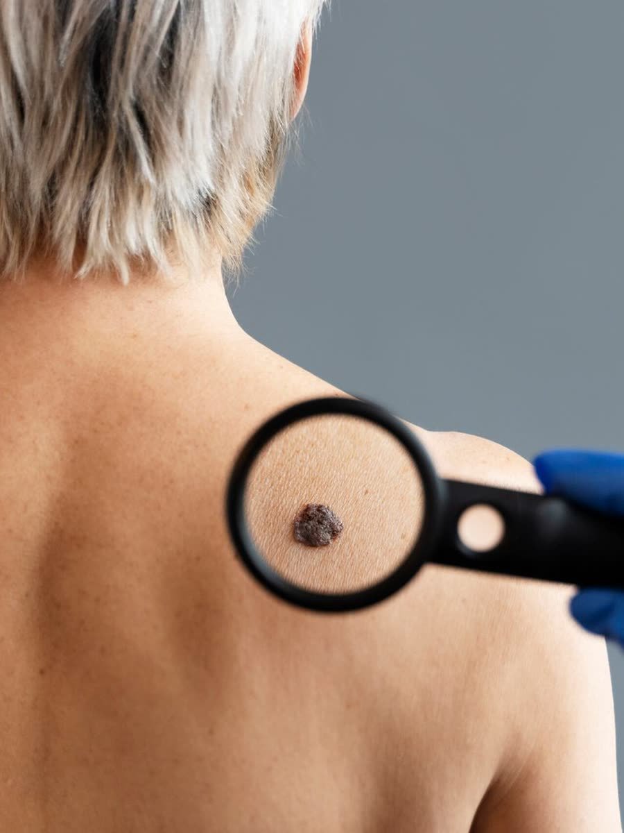
Skin Cancer
Skin cancer is the most common type of cancer in the world and, unfortunately, it is still widely neglected. The main risk factor is sun exposure, and even today we see people out in the sun during the hours of highest radiation, without sunscreen, without protective clothing and, worse still, exposing children.
Children, especially up to 5 years of age, need extra care. A sunburn in early childhood greatly increases the chance of developing skin cancer in adulthood. The good news is that, when detected early, skin cancer has high cure rates, which makes prevention and early diagnosis essential.
The three types of skin cancer
Basal Cell Carcinoma
The most common and the least aggressive. It grows slowly, rarely causes metastasis, and has cure rates close to 100% when diagnosed early.
Squamous Cell Carcinoma
Less common than basal cell carcinoma, but more aggressive and with a greater potential to spread to the lymph nodes and other organs. Early diagnosis is essential.
Melanoma
The most dangerous type. It can cause metastasis and be fatal if not treated in time. Despite being more aggressive, it has a high cure rate when detected at an early stage.
Basal Cell Carcinoma (BCC)
This is the most common type of skin cancer and, fortunately, also the least malignant. It usually grows slowly and rarely spreads to other parts of the body. It is highly treatable when diagnosed early.
Risk factors
- Prolonged sun exposure, especially on frequently exposed areas such as the face, neck, and arms.
- Fair skin and a family history of skin cancer.
Signs and symptoms
- A lesion or spot on the skin that is pink, red, or brown.
- It may appear as a mole or a sore that does not heal.
- It often bleeds easily and forms a crust.
- It is very common on the nose and other areas of the face.
Severity
- Considered the least aggressive of the skin cancers: it grows slowly and rarely spreads to other organs.
- When diagnosed early, treatment is highly effective, with cure rates of almost 100%.
Treatment
- The main treatment is surgical removal of the cancerous lesion.
- For the superficial type, other options include photodynamic therapy, cryotherapy (extreme cold), laser excision, and topical medications such as imiquimod.
Prevention
- Daily sun protection with an appropriate SPF and clothing with UV protection.
- Regular exams with a dermatologist to evaluate any suspicious lesion.
Although basal cell carcinoma is generally the least worrisome of the skin cancers, any suspicious sign or lesion should be evaluated. Early detection and timely treatment are essential to ensure a cure and avoid complications.
Squamous Cell Carcinoma (SCC)
Squamous cell carcinoma is less common than basal cell carcinoma, but it can be more aggressive and has a greater potential to spread. Early diagnosis is essential for successful treatment.
Risk factors
- Prolonged, chronic sun exposure throughout life, as well as severe sunburns.
- A history of exposure to carcinogenic substances, such as arsenic.
- A weakened immune system: transplant patients and people with HIV infection are at higher risk.
Signs and symptoms
- A rough, red, scaly lesion that may be painful.
- It may start as a mole, sore, or bump that does not heal or that grows quickly.
- Irregular borders, with color ranging from pink to red or brown.
Severity
- More aggressive than basal cell carcinoma, but less aggressive than melanoma.
- If left untreated, it can spread to the lymph nodes and, occasionally, to other organs. When diagnosed and treated early, cure rates are high.
Treatment
- Surgical removal of the cancerous lesion.
- In advanced cases or when it has spread, radiation therapy, chemotherapy, or targeted therapy may be needed.
Prevention
- Regular use of sunscreen, proper sun protection, and periodic skin exams.
- Watch for lesions that do not heal, grow quickly, or change in appearance, and see a dermatologist.
Early diagnosis and timely treatment remain essential for successful treatment. Prevention and self-care for your skin play an important role in reducing the risk.
Melanoma
Melanoma is the most dangerous type of skin cancer and can cause metastasis, spreading to other organs. Detecting it early is crucial, because timely treatment significantly increases the chances of a cure. Although it is more aggressive, it also has a high cure rate when identified at an early stage.
Risk factors
- Sun exposure: severe sunburns, especially in early childhood, increase the risk of melanoma in adulthood.
- Many skin growths (moles or nevi) or a family history of melanoma.
- Exposure to tanning beds and artificial ultraviolet radiation.
Signs and symptoms
- A mole or mark on the skin that changes in size, shape, color, or texture.
- It may be asymmetrical, have irregular borders, several colors, and a diameter larger than 6 mm (the ABCD rule), and it may evolve over time.
- It usually causes no symptoms: it grows silently, which makes regular follow-up even more important.
- Detection may not be easy on clinical examination alone and may require specific exams such as dermoscopy and digital body mapping.
Severity
- Considered an aggressive cancer because of its potential to spread quickly to the lymph nodes and distant organs, which makes it potentially lethal.
- When diagnosed at early stages, treatment is highly effective, with very high cure rates.
Treatment
- It always involves surgical removal of the lesion and the surrounding tissue. The surgeon must have a solid command of surgical margins, both in the biopsy and in the wide local excision. The surgery is done in two stages: first with a small margin to confirm or rule out the diagnosis; then, once melanoma is confirmed, a wide local excision is performed (margins from 0.5 cm to 2 cm, according to the Breslow thickness, that is, the depth of the lesion reported by the biopsy).
- In advanced cases or when it has spread, immunotherapy, targeted therapy, or chemotherapy may be needed.
- Invasive melanomas require blood tests and imaging tests, with joint follow-up by an oncologist.
Prevention
- Proper sun protection, avoiding tanning beds, and checking your skin growths regularly.
- Regular self-exams and medical consultations to evaluate suspicious moles. Knowing your moles (what is new and what has changed) is essential for the medical evaluation.
Any suspicious change in the skin, especially in moles you already know, should be evaluated by a dermatologist right away.
The ABCDE rule for identifying suspicious lesions
The ABCDE rule is a simple and effective guide for identifying moles or spots that deserve medical attention. Look at any skin growth using these criteria:
A: Asymmetry
One half of the mole does not match the other. Normal moles tend to be symmetrical.
B: Borders
Irregular, jagged, or poorly defined borders, or borders that seem to “bleed” into the surrounding skin.
C: Color
Color variations within the same lesion: shades of brown, black, red, white, or blue.
D: Diameter
Lesions with a diameter larger than 6 mm (roughly the size of a pencil eraser) are more suspicious.
E: Evolution
Any recent change in size, shape, color, texture, or symptoms (itching, bleeding) should be evaluated.
Who has a higher risk of developing skin cancer
Excessive sun exposure
The main risk factor for all types. UV radiation accumulates over a lifetime and damages the DNA of skin cells.
Childhood sunburns
A severe sunburn in the first years of life significantly increases the risk of skin cancer in adulthood.
Fair skin and light eyes
People with lighter skin phototypes produce less protective melanin and are more vulnerable to damage from UV radiation.
Family history
Having relatives with skin cancer or melanoma raises your individual risk, and it is something you should tell your dermatologist.
Many moles (nevi)
A large number of skin growths, especially atypical ones, increases the chance of melanoma and calls for regular follow-up.
Immunosuppression
Transplant patients and people with diseases that compromise the immune system have a higher risk, especially of squamous cell carcinoma.
Tanning beds
Artificial ultraviolet radiation is a proven risk factor for all types of skin cancer.
Exposure to chemicals
Chronic contact with arsenic and other carcinogenic agents raises the risk, especially of squamous cell carcinoma.
How to reduce the risk of skin cancer
Prevention is the best and most effective defense. Adopting protective habits early on can significantly reduce the risk of developing skin cancer over a lifetime.
Daily sun protection
Use sunscreen with an appropriate SPF whenever you are outdoors, even on cloudy days. Reapplying every 2 hours is essential during prolonged exposure.
Protective clothing and accessories
Long-sleeved shirts with UV protection, wide-brimmed hats, and sunglasses with UVA and UVB protection complement the barrier against radiation.
Avoid the sun at peak hours
Stay in the shade between 10 a.m. and 4 p.m., when the sun’s rays are most intense and have the greatest potential for cell damage.
Protect children
Children up to 5 years old need extra attention. Avoid direct sun exposure and apply sunscreen formulated for their age group.
Skin self-exam
Regularly check all of your skin for spots, moles, or lesions that are new or have changed. See a dermatologist if you notice any change.
Avoid tanning beds
Artificial UV radiation is a proven carcinogen, and there is no safe dose for indoor tanning.
Remember: prevention is the cure. By adopting these habits, you significantly reduce the risk of one of the most common (and most preventable) diseases in the world.
Digital Body Mapping: technology in the service of early detection
Skin cancer is the most common type of cancer in the world, but early detection is essential for effective treatment. Digital body mapping is an important innovation in prevention and early diagnosis, offering a highly precise and noninvasive approach. Through a system that organizes and magnifies the lesions, we considerably increase the chances of identifying changes at an early stage.
At the clinic, we use Fotofinder, equipment used internationally for digital body mapping and computerized dermoscopy.
How Digital Body Mapping works
- Body imaging: The patient is photographed from multiple angles by a high-resolution camera, including areas that are hard to self-examine.
- Evaluation with a dermatoscope: The dermatologist manually evaluates the moles with a dermatoscope and selects those that should be analyzed in greater detail with Fotofinder.
- Comparative image analysis: The software compares the photographs over time, identifying subtle changes in moles and other marks that may indicate a developing cancer.
- Medical report: The dermatologist interprets the results and provides a detailed report. If a suspicious lesion is identified, a biopsy or additional treatment may be recommended. The software displays and compares the images, but the interpretation and the course of action are always the physician’s.
Advantages of Digital Body Mapping
- Early detection: It makes it possible to identify lesions before they become visible to the naked eye.
- Long-term monitoring: It tracks moles over time, picking up subtle changes that could go unnoticed.
- Noninvasive: It does not require routine biopsies, reducing discomfort for the patient.
- High precision: It reduces false positives and false negatives; diagnostic confirmation, when needed, is made by biopsy.
- Convenience: A quick procedure with no recovery time that fits into the dermatology consultation.
Digital body mapping is especially indicated for patients with many nevi, a personal or family history of melanoma, or other risk factors. Talk to your dermatologist to find out whether this exam is right for your case.
Severity varies: early diagnosis saves lives
The severity of skin cancer varies according to the type and stage of the disease. Basal cell carcinoma and squamous cell carcinoma, when diagnosed at early stages, generally have very high survival rates. Melanoma is more aggressive and can be fatal if not treated early, which is why any sign of skin cancer should be evaluated by a physician without delay.
Stay informed, take care of your skin, and see a health professional whenever you notice any change. Together, prevention and early diagnosis are the best strategy against skin cancer.
Digital Body Mapping
In Brazil, retirees and pensioners with skin cancer do not pay income tax
Few people know this, but Brazil’s income tax law (Lei 7.713/1988, art. 6º, inciso XIV) exempts the retirement pension, military retirement pay, and survivor’s pension of people who have a malignant neoplasm, and skin cancer falls into this category. The right applies even when the diagnosis is old or the treatment has already ended, and the amounts paid over the last five years can be claimed back. The exemption applies to this income, not to salaries or other earnings.
Taxes are not our field. Anyone who wants to understand whether they have this right and how to apply for it can find an explanation of the criteria and the step-by-step process at Pede Ajuda, a team of lawyers specializing in income tax exemption for serious illness.
Skin cancer prevention and treatment in Barra da Tijuca and Ipanema
Skin cancer prevention and treatment are available at both Drummond Dermato locations in Rio de Janeiro. In Barra da Tijuca, the clinic is at Av. das Américas, 3200, sala 112. In Ipanema, at Rua Visconde de Pirajá, 540, sala 314. At both, the treatment is performed by dermatologists from our team, after a consultation that evaluates the skin and defines the protocol.
Where to get mole mapping in Rio de Janeiro?
At Drummond Dermato, in Barra da Tijuca or Ipanema. Book your consultation via WhatsApp and choose the location closest to you.
Prevention and early diagnosis save lives
Book your dermatology consultation in Ipanema or Barra da Tijuca and care for the health of your skin with people who know it well.
Book via WhatsApp